Artificial Intelligence and Machine Learning may sound like terms used strictly by computer engineers, but recent experiments by pathologists at the James A. Haley Veterans’ Hospital have shown they can also be used to help diagnose cancer, and possibly other health issues.
The investigation looked at two AI platforms to see if they could use images of tissue samples to differentiate between normal or benign cells and cancerous cells. Those platforms are Apple Create ML and Google AutoML. They also wanted to see if the programs could tell the difference between three different types of cancer.
According to Dr. Andrew Borkowski, AI is anything a computer can do that is normally a task that requires human intelligence. Borkowski, JAHVH chief of Molecular Diagnostics Section, is one of the pathologists participating in the project. Machine Learning is a branch of AI where the computer learns from examples in order to make predictions.
“A pretty basic version of this is that we try to teach the computer to differentiate between the cancer image and the benign tissue image,” Borkowski said. “We did it for two of the most common cancers that are in our Veteran population, which are colon cancer and lung cancer.”
Accuracy of 90 per cent or higher
Using a microscope with an attached camera, Borkowski and his team photographed 1,250 different tissue samples mounted on slides and removed any personally identifiable information from them. Doctors collected samples during biopsies and other medical procedures. The samples included benign tissue and slides that indicated three different types of cancer. Those cancers are colon adenocarcinoma, lung squamous cell carcinoma and lung adenocarcinoma.
The doctors then input about 200 slides from the different classes into the AI computer programs to help them learn the differences between each type of cancer and healthy tissue. Fifty other slides from each class, unknown to AI network, were then used to test the systems, with surprising results.
“We did six experiments, and with most of the experiments we achieved an accuracy rate of 90 percent or higher,” Borkowski said.
Dr. Stephen Mastorides added that the computer went beyond just identifying whether a slide was cancer or healthy tissue. Mastorides is chief of the JAHVH Pathology and Laboratory Service.
“For distinguishing between benign tissue and cancer, between colon cancer and lung cancer, and between the two types of lung cancer,” Mastorides said. “The accuracy was over 90 percent for each of those things.”
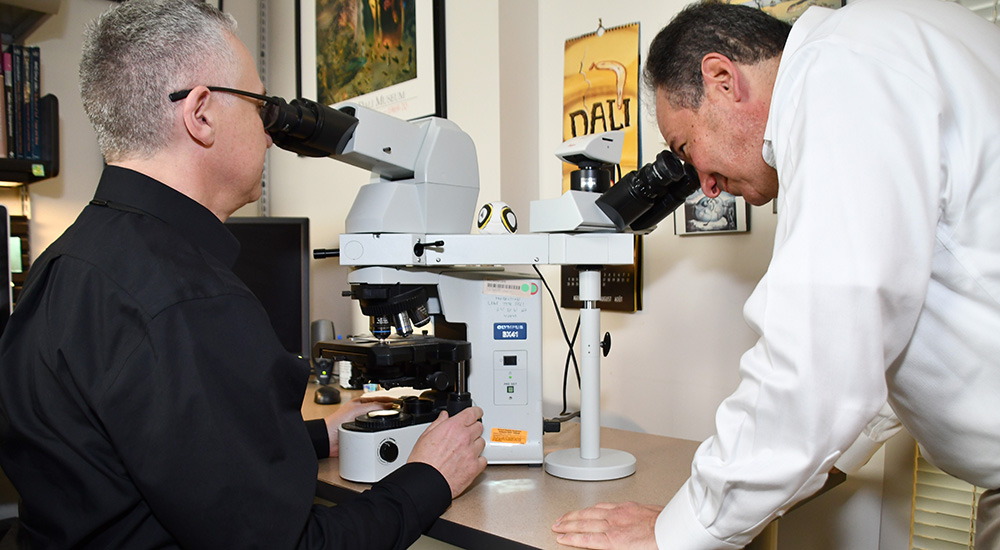
From left, JAHVH pathologists Dr. Andrew Borkowski and Dr. Stephen Mastorides examine tissue sample slides under a microscope. The microscope also captures images that can train the computers.
They also tested whether the computer could tell if a specific mutation was evident in the colon cancer slides. The computer successfully identified the KRAS mutation about 70 percent of the time. That is still good. Pathologists typically need to do a molecular test in the lab to identify this particular mutation. Both computer platforms performed about the same in all the tests.
Important because of different treatment regimens
Borkowski said it’s important to differentiate between the different cancers because each requires a different treatment regimen.
The test team is not settling for just these forms of cancer, though. It already is in the process of expanded AI testing with Dr. Narayan Viswanadhan from the radiology department. They are now training computers with images that include brain hemorrhages. That could eventually play an important role in the emergency department.
“Right now, we’re going to be running a model and training the AI to see if it can diagnose the hemorrhages, which would be phenomenal,” Borkowski said. “Let’s say a patient comes to the emergency room and there’s no radiologist available right away. You can capture the (MRI) image, run it through the program and it says ‘Hey, here’s a brain hemorrhage, let’s do something.’”
While the accuracy of the AI diagnoses was very good, the intent is not to eventually replace doctors. Instead, the program gives them tools to improve quality and increase productivity, Borkowski explained.
“Our ultimate goal would be to create programs that can be rolled out in the entire VA system so that pathologists who are working solo, or maybe there are two pathologists in some small VAs, would have the benefit of having something that is helping them become more productive, help them prioritize the workload and improve quality,” Borkowski said. “We see a huge future for AI in pathology, radiology, dermatology – any medical specialty that is dealing with images.”
Note: The results of the AI experiment were recently published in Federal Practitioner magazine and can be seen at https://www.mdedge.com/fedprac/article/209325/health-policy/comparing-artificial-intelligence-platforms-histopathologic.
Topics in this story
More Stories
Study underscores important role COVID vaccination can have in protecting Veterans from infection and reducing long-term health consequences
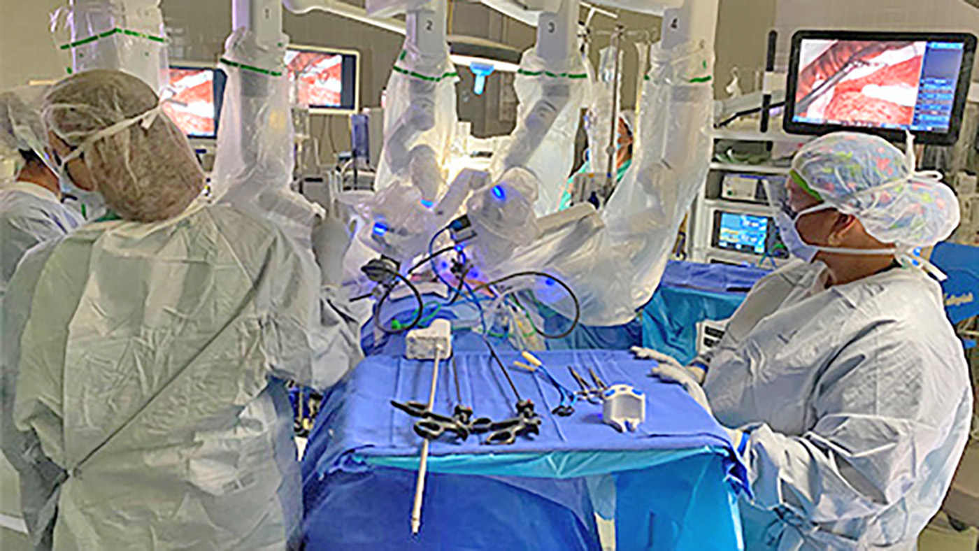
Columbia VA’s robotic surgery teams completed their 800th robotic surgery and are on schedule to hit 1,000 by the end of the year.

In a decentralized clinical trial, Veterans can participate from their own homes or local VA instead of having to travel to a research site.