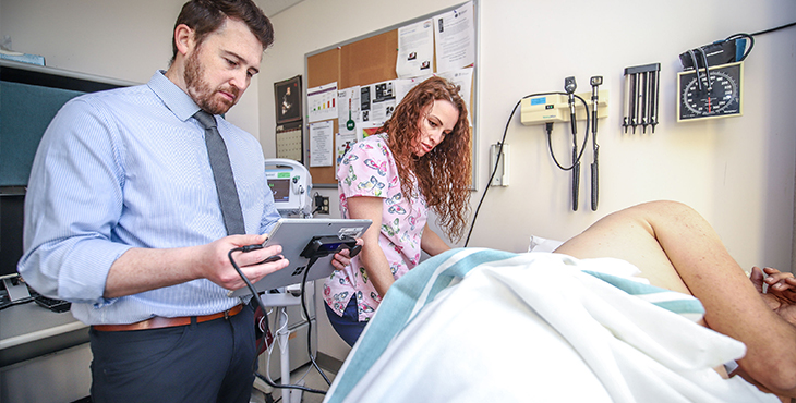
Above: Researcher Dr. Matthew Peterson and nurse Trina Dickert use an experimental new automated system for measuring bedsores, or pressure ulcers—a common problem for Veterans with spinal cord injury, as well as other patient populations. (Photo by Dan Henry)
VA researchers are developing a new instrument for measuring bedsores, using 3D cameras, tablet computers, and algorithms, to help treat the problem in Veterans with spinal cord injuries.
Investigators at the James A. Haley Veterans’ Hospital in Tampa, Florida, are testing an innovative computer-based system designed to more accurately measure bedsores, also known as pressure ulcers. Accurate measurement of the wound, followed by optimal treatment, is key to preventing it from worsening.
Pressure ulcers are painful injuries to the skin and underlying tissue resulting from prolonged pressure on the skin. People with spinal cord injuries are at high risk for pressure ulcers due to immobility and other risk factors. The wounds most often develop around bony areas of the body, such as the heels, ankles, hips, and tailbone.
“This digital technique, which basically captures the whole image rather than just parts of it, is going to provide greater accuracy in measuring the wound.”
In general, the worse the pressure ulcer, the more advanced the care needed for it to heal. However, assessment of the wound has traditionally been based on a manual process that can lead to treatment challenges.
“That type of measurement can’t accurately account for small improvements or deterioration over the entire surface area, and doesn’t consider changes in the wound’s depth,” says Dr. Matthew Peterson, a biomedical engineer who is leading the testing of the computer-based system. “Accurate pressure ulcer measurement is vital for clinicians to accurately assess the severity of the wound and the degree of tissue damage, and to determine whether current treatment strategies are effective.
“If the size of the pressure ulcer has been shrinking over time, the wound is healing. But if the size stays the same or increases, the treatment may not be effective and may need to be modified. Therefore, effective treatment depends on reliable and valid measurements of the pressure ulcers. Intervening early to prevent them from worsening is critical for improving patient outcomes and reducing costs.”
Peterson is testing a technology that’s designed to be more objective and reliable than the manual system in current use. His system captures 3D images with a camera mounted to a tablet. An algorithm uses the color, geometry, and formation of the wound to assess its length, width, and depth.
“This digital technique, which basically captures the whole image rather than just parts of it, is going to provide greater accuracy in measuring the wound,” Peterson says. “We’re capturing the full perimeter and diameter, rather than just having one measurement for length, one measurement for width, and one measurement for depth. Essentially, we aim to determine the size of the wound based on its entire features.”
He explains, for example, that because pressure ulcers can be shaped irregularly, the spot where the depth is marked in a manual system may not be the deepest point in the wound. His instrument is designed to more accurately mark that point.
Read more of this story at VA Research Currents.
To learn more about VA research on spinal cord injury, visit the SCI topic page on the VA Research website.
Topics in this story
More Stories
In a new series that highlights advancements in VA health care, VA researchers and clinicians are appearing on a Veteran-themed media platform—Wreaths Across America Radio—to tout their critical work.
Recently published findings from the VA Disrupted Care National Project [...]
Diverse representation of women in health care research allows MVP to make discoveries for women’s health


